Neisseria meningitidis — Meningococcus

Core concept: Neisseria meningitidis is a human-restricted, encapsulated, Gram-negative diplococcus that commonly colonizes the nasopharynx but can occasionally invade the bloodstream and CNS, producing meningitis, meningococcemia and fulminant septic shock.
1. Why meningococcus matters
Think of N. meningitidis as:
Carrier state → nasopharyngeal invasion → bacteremia → endothelial injury → meningitis / septicemia
The two major invasive syndromes are:
Meningococcal meningitis
Meningococcemia/septicemia
It can progress extraordinarily rapidly, and mortality remains substantial even with treatment. CDC currently estimates approximately 10–15% mortality, while about 1 in 5 survivors may have long-term sequelae.
2. Taxonomy

Common name
Meningococcus
Important related species:
N. gonorrhoeae
N. meningitidis
N. lactamica
N. sicca
N. subflava
N. mucosa
The big clinical distinction
Meningococcus → meningitis + septicemia
Gonococcus → urethritis/cervicitis + genital infection
3. Morphology
Classic appearance
Gram-negative, kidney/coffee-bean-shaped diplococci
They are frequently seen:
Intracellularly within neutrophils
and extracellularly in clinical specimens.
Important morphological features
Gram-negative
Diplococci
Adjacent flattened surfaces
Kidney/coffee-bean appearance
Often intracellular in PMNs
Encapsulated
Non-spore-forming
Non-motile
Exam image in words
Gram-negative diplococci + PMNs + CSF = think N. meningitidis.
However:
Gram-negative diplococci in CSF are strongly suggestive but not by themselves confirmatory.
4. Culture characteristics
Meningococcus is relatively fastidious.
Growth requirements
It grows well on:
Chocolate agar
Blood agar
Selective media such as Thayer–Martin / modified Thayer–Martin
Optimal incubation:
Approximately 35–37°C
Increased CO₂ atmosphere
Moist environment
Colonies
Typical colonies are:

5. Important biochemical reactions

Memory trick
MeninGitidis- Maltose+Glucose
Gonococcus-Glucose
Historically, carbohydrate utilization was an important identification method; modern laboratories may use MALDI-TOF, molecular methods and other validated identification systems.
6. Oxidase test
Principle
Neisseria possesses cytochrome c oxidase.
Therefore:
N. meningitidis = oxidase positive
A positive oxidase reaction produces a rapid purple color.
Exam sequence
Gram-negative diplococcus → oxidase positive → glucose + maltose → meningococcus
7. Capsule — the superstar virulence factor ⭐
The capsule is one of the most important determinants of invasive disease.
It:
Prevents phagocytosis
Helps resist complement-mediated killing
Promotes survival in bloodstream
Facilitates invasive disease
Forms the basis of serogroup classification
Serogroups
The clinically important meningococcal serogroups include:
A
B
C
W
X
Y
These six account for >95% of meningococcal disease globally, although their relative importance varies geographically and temporally.
8. Serogroup ≠ serotype
This distinction is extremely important.
Serogroup
Determined primarily by:
Capsular polysaccharide
Serotype
Determined by:
Outer-membrane proteins
So:
Capsule → serogroup
Outer membrane proteins → serotype/subtyping
9. Virulence factors
Think of meningococcal virulence as a multi-layered survival system.
Major virulence factors
Capsule
LOS endotoxin
Type IV pili
Opa proteins
Opc protein
IgA protease
Factor H-binding protein
Outer membrane proteins
Iron acquisition systems
10. Capsule
Functions
The capsule:
↓ phagocytosis
↓ complement-mediated killing
↑ bloodstream survival
Encapsulated organisms are therefore much more capable of producing invasive disease.
11. LOS — the endotoxin
Meningococcus possesses:
Lipooligosaccharide (LOS)
Unlike enteric Gram-negative bacteria, meningococci characteristically lack the long O-polysaccharide side chain of classical LPS.
So:
Meningococcus
LOS
Enterobacterales
LPS
Why LOS is dangerous
LOS stimulates massive inflammatory responses through innate immune pathways.
This can produce:
TNF-α
IL-1
IL-6
endothelial activation
vascular leakage
vasodilatation
disseminated intravascular coagulation
shock
This explains the terrifying clinical picture of:
Meningococcemia → purpura → DIC → shock → multiorgan failure
12. Type IV pili
Pili are essential for:
Initial colonization
They help meningococci:
Attach to nasopharyngeal epithelial cells
Resist mechanical clearance
Interact with host cells
Initiate invasion
They also undergo antigenic variation, helping immune evasion.
13. Opa and Opc proteins
These are outer-membrane adhesins.
They facilitate:
close interaction with host epithelial/endothelial cells
and contribute to cellular invasion.
14. IgA protease
The organism colonizes a mucosal surface bathed in IgA.
It produces:
IgA protease
which cleaves secretory IgA and facilitates mucosal colonization.
15. Factor H-binding protein — high-yield modern concept
One of the particularly elegant immune-evasion mechanisms is:
fHbp → binds human factor H → downregulates alternative complement pathway → increased bacterial survival
This interaction helps meningococcus evade complement-mediated killing.
And this protein isn’t merely biologically interesting:
fHbp is also an important antigen used in serogroup B meningococcal vaccine strategies.
16. Iron acquisition
Iron is essential for bacterial growth, but free iron is tightly restricted in humans.
Meningococcus therefore possesses systems to acquire iron from host sources, including:
Transferrin
Lactoferrin
Hemoglobin/heme-associated sources
This becomes particularly important during invasive infection.
17. Pathogenesis — the story you should remember
Step 1 — Colonization
Transmission occurs through:
Respiratory droplets / respiratory secretions during close contact
The organism colonizes the nasopharynx.
Most carriers remain asymptomatic.
Step 2 — Adhesion
Pili and outer-membrane adhesins attach meningococcus to nasopharyngeal epithelium.
↓
Step 3 — Translocation
The organism can cross the epithelial barrier.
↓
Step 4 — Bacteremia
Meningococci enter the bloodstream.
↓
Step 5 — Survival
Capsule + complement evasion mechanisms allow survival.
↓
Step 6 — Endothelial interaction
Meningococci interact with vascular endothelial cells.
↓
Step 7 — Massive inflammatory response
LOS triggers intense cytokine and inflammatory responses.
↓
Step 8 — Vascular injury
↓
Step 9 — DIC + shock
↓
Step 10 — CNS invasion
Crossing the blood–brain barrier allows:
Meningococcal meningitis

18. Carrier state
This is a crucial epidemiological concept.
The organism can exist harmlessly in the:
Nasopharynx
without causing disease.
Therefore:
Colonization ≠ disease
Only a small proportion of colonized individuals develop invasive disease.
19. Transmission
Transmission occurs primarily through:
Respiratory droplets
Salivary/oral secretions
Close or prolonged contact
Examples:
Household contact
Kissing
Crowded living conditions
Dormitories
Military barracks
Certain mass gatherings
The organism is adapted to humans and does not survive well outside the human host.
20. Incubation period
Approximately:
2–10 days
Usually:
3–4 days
21. Risk factors for invasive disease
Important risk factors include:
Host factors
Complement deficiency, especially terminal complement pathway defects
Functional/anatomic asplenia
HIV infection
Certain immunodeficiencies
Complement inhibitor therapy
Epidemiological factors
Close household contact
Crowding
Adolescence/young adulthood
Certain geographic regions
Mass gatherings
Environmental factors
Respiratory infections and smoking exposure can facilitate acquisition/invasion.
22. Clinical spectrum
Think:
Meningitis + bloodstream infection + focal disease
A. Meningococcal meningitis
Classic triad:
Fever + headache + neck stiffness
Other manifestations:
Photophobia
Vomiting
Altered consciousness
Seizures
Confusion
Coma
WHO emphasizes that meningococcal meningitis is among the major causes of acute bacterial meningitis globally.
23. Meningococcemia
This is the bloodstream infection form.
Characteristic findings:
Fever
Toxic appearance
Hypotension
Petechiae
Purpura
DIC
Shock
Multiorgan failure
Classic rash
Petechial/purpuric, non-blanching rash
This should immediately make you think of:
Meningococcemia
24. Waterhouse–Friderichsen syndrome
One of the most important exam associations.
Meningococcemia
↓
Endotoxin-mediated shock
↓
DIC
↓
Bilateral adrenal hemorrhage
↓
Acute adrenal insufficiency
= Waterhouse–Friderichsen syndrome
Clinical picture may include:
Profound shock
Purpura
DIC
Adrenal failure
Multiorgan dysfunction
25. Meningococcal pneumonia
Meningococcus can cause pneumonia, particularly in certain adults and outbreaks.
It may present with:
Fever
Cough
Chest pain
Respiratory symptoms
But compared with meningitis and meningococcemia, pneumonia is a less dominant manifestation.
26. Other focal infections
Rarely:
Septic arthritis
Pericarditis
Endocarditis
Conjunctivitis
Otitis
Urethritis
So don’t make the mistake:
“N. meningitidis only causes meningitis.”
27. Laboratory diagnosis
Patient with suspected meningococcal meningitis
Patient
↓
CSF + blood
↓
Gram stain
↓
Culture
↓
Identification
↓
Molecular confirmation if required
↓
Serogrouping
↓
AST
28. Specimens
Depending on presentation:
Meningitis
CSF
Blood
Meningococcemia
Blood
Skin lesion/purpuric lesion material where appropriate
Other invasive disease
Normally sterile site specimens
CDC recognizes culture or validated PCR from normally sterile sites such as CSF or blood as diagnostic approaches.
29. CSF findings
Typical bacterial meningitis pattern:
Appearance
Turbid
Cloudy
Cells
Predominantly:
Neutrophils
Protein
↑
Glucose
↓
CSF/serum glucose ratio
↓
Opening pressure
↑
30. CSF Gram stain
Classic:
Intracellular Gram-negative diplococci
This is one of the most useful rapid clues.
But:
Gram stain = presumptive
Culture/PCR = confirmation
31. Culture
Culture remains an important reference/gold-standard diagnostic method because it provides an isolate that can be used for:
Identification
Serogrouping
Antimicrobial susceptibility testing
Molecular epidemiology
Whole-genome sequencing
CDC notes that culture has very high specificity but sensitivity decreases with improper specimen handling and prior antibiotic exposure.
32. Chocolate agar
Because meningococcus is fastidious:
Chocolate agar is an important primary isolation medium.
Blood agar may also support growth.
33. Thayer–Martin medium
A selective medium for pathogenic Neisseria.
It contains antimicrobial agents that suppress competing flora.
Classically associated selective components include:
Vancomycin
Colistin
Nystatin
Some formulations also contain additional agents such as trimethoprim.
Purpose
Suppress contaminating Gram-positive organisms and many Gram-negative rods while allowing pathogenic Neisseria to grow.
34. Identification algorithm
Step 1
Gram-negative diplococcus
↓
Step 2
Oxidase positive
↓
Step 3
Glucose + maltose positive
↓
Step 4
Confirm with:
MALDI-TOF
Molecular assays
Serogrouping
Other validated identification systems
35. MALDI-TOF
Modern clinical laboratories increasingly use:
MALDI-TOF MS
for rapid species identification.
It can distinguish N. meningitidis from other Neisseria species when the laboratory’s validated database/workflow supports it.
36. PCR
PCR is particularly useful when:
Antibiotics were given before specimen collection.
Why?
Because antibiotics can sterilize CSF/blood cultures while bacterial nucleic acid may remain detectable.
CDC specifically notes that PCR can detect meningococcus when culture is negative after prior antibiotic exposure.
37. Serogroup identification
Once N. meningitidis is confirmed:
Determine the serogroup.
Methods include:
Slide agglutination
Real-time PCR
Other validated molecular methods
Serogroup information is extremely important for:
Surveillance
Outbreak investigation
Vaccine strategy
Epidemiology
38. Antimicrobial susceptibility testing
AST matters because resistance is increasingly relevant.
Important resistance mechanisms include:
Altered penicillin-binding proteins
β-lactamase production
Fluoroquinolone resistance
A key current public-health issue is:
Ciprofloxacin-resistant N. meningitidis
which can compromise ciprofloxacin chemoprophylaxis.
39. Treatment
Suspected meningococcal disease
Do not wait for microbiological confirmation before starting antibiotics.
Current CDC guidance recommends:
Ceftriaxone or cefotaxime
as empiric therapy for suspected meningococcal disease.
Once confirmed
A third-generation cephalosporin can generally be continued.
If susceptibility is established, therapy may be narrowed to:
Penicillin G or ampicillin
according to susceptibility and local guidance.
40. Important treatment pearl
Treating disease ≠ eradicating carriage
Some systemic antibiotic regimens may successfully treat invasive disease but may not reliably eradicate nasopharyngeal carriage.
If ceftriaxone/cefotaxime is not used, additional carriage-eradication therapy may be required before discharge.
41. Chemoprophylaxis
This is not treatment of the patient.
It is for:
Close contacts
The purpose is:
Eradicate nasopharyngeal carriage → prevent secondary cases
CDC recommends prophylaxis for close contacts regardless of their vaccination status.
42. Who needs prophylaxis?
Typical high-risk close contacts include:
Household contacts
Intimate contacts
Persons directly exposed to oral/nasal secretions
Certain healthcare workers with unprotected exposure to respiratory/oral secretions
Not routinely indicated for:
Simply being in the same hospital/room without significant exposure to oral/respiratory secretions.
Also:
Asymptomatic carriers without known close contact do not routinely require treatment.
43. Chemoprophylaxis drugs
Common options:
1. Rifampicin
Usually:
2-day regimen
2. Ceftriaxone
Single IM dose
3. Ciprofloxacin
Single oral dose, provided local susceptibility supports its use.
4. Azithromycin
Not routinely first-line, but may be considered in settings with sustained ciprofloxacin resistance.
44. Timing of prophylaxis
The sooner the better.
Ideally:
Within 24 hours of identifying the index case/exposure
Benefit decreases substantially when given late.
45. Vaccines
Vaccines target important meningococcal serogroups.
Major vaccine approaches include:
MenACWY
Protects against:
A
C
W
Y
MenB
Targets:
Serogroup B
MenABCWY
Combination vaccine covering:
A + B + C + W + Y
Current vaccine recommendations vary by country, age and risk factors.
46. Why serogroup B is different
This is a beautiful microbiology point.
The capsule of serogroup B contains:
α-2,8-linked polysialic acid
which resembles human neural-cell surface structures.
Therefore, developing a conventional capsule-polysaccharide vaccine against MenB is problematic.
This led to development of:
Protein-based MenB vaccines
using antigens such as fHbp and other surface proteins.
47. Global epidemiology
Meningococcal disease occurs worldwide.
The highest historic burden of meningococcal meningitis has been associated with the:
African meningitis belt
stretching across sub-Saharan Africa.
Historically, serogroup A was a major epidemic cause there; widespread deployment of MenA conjugate vaccination dramatically changed the epidemiology. WHO also notes the introduction of a pentavalent Men5CV vaccine targeting A, C, W, Y and X in the African meningitis belt.
48. Epidemics — why meningococcus is special
Meningococcus has substantial epidemic potential.
Risk increases with:
Crowding
Close interpersonal contact
Mass gatherings
Military camps
Refugee/displacement settings
Certain geographic environments
49. The complement connection
This is one of the most important immunology associations.
Terminal complement deficiency
Deficiency of:
C5 → C6 → C7 → C8 → C9
impairs:
Membrane attack complex (MAC) formation
↓
Poor killing of Neisseria
↓
Recurrent meningococcal infections
Exam pearl
Recurrent Neisseria infections → think terminal complement deficiency.
Also remember increased risk with certain complement-inhibiting therapies.
50. Meningococcus vs Gonococcus

Memory:
Meningococcus = Maltose + Meningitis
51. Meningococcus vs pneumococcus
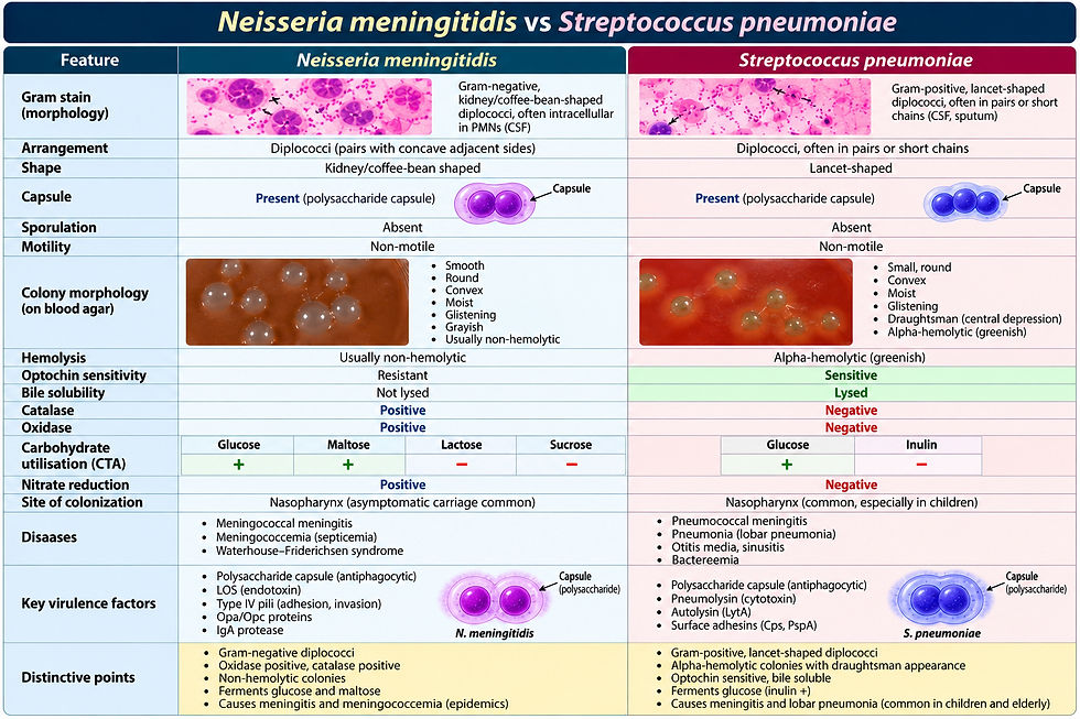
Fast exam distinction
Gram-negative diplococci → meningococcus
Gram-positive lancet diplococci → pneumococcus
52. Meningococcal meningitis — diagnostic workflow
SUSPECT MENINGOCOCCAL MENINGITIS
↓
Blood + CSF
↓
CSF Gram stain
↓
G− intracellular diplococci
↓
Start treatment
↓
Culture on appropriate media
↓
Oxidase-positive Neisseria
↓
Identification confirmation
↓
PCR if indicated
↓
Serogroup determination
↓
AST
↓
Epidemiology + contact tracing
↓
Chemoprophylaxis contacts
53. The meningococcal sepsis pathway
Nasopharyngeal carriage
↓
Mucosal invasion
↓
Bacteremia
↓
Capsule-mediated survival
↓
Endothelial interaction
↓
LOS-mediated inflammation
↓
Cytokine storm
↓
Vascular injury
↓
DIC
↓
Purpura
↓
Shock
↓
Multiorgan failure
54. The “one organism → five mechanisms” framework
For rapid revision:
CAPSULE
→ antiphagocytic
PILI
→ adhesion/colonization
IgA PROTEASE
→ mucosal immune evasion
LOS
→ endotoxin/sepsis
fHBP
→ complement evasion
That five-part framework captures a huge amount of meningococcal biology.
55. High-yield clinical associations ⭐⭐⭐
Finding
Think
G− diplococci in CSF
N. meningitidis
Meningitis + petechiae
Meningococcemia
Purpura + shock
Fulminant meningococcemia
Bilateral adrenal hemorrhage
Waterhouse–Friderichsen
Recurrent Neisseria infection
Terminal complement deficiency
Glucose + maltose
N. meningitidis
Glucose only
N. gonorrhoeae
Capsule
Major virulence determinant
LOS
Endotoxin
Type IV pili
Adhesion
IgA protease
Mucosal survival
fHbp
Complement evasion + MenB vaccine antigen
Close contact
Chemoprophylaxis
Ciprofloxacin resistance
Consider alternative prophylaxis
56. Classic examination traps
❌ “All nasopharyngeal carriers need antibiotics.”
False.
Asymptomatic carriage alone is not an indication for routine treatment.
❌ “Gram-negative diplococci in CSF is definitive.”
False.
It is highly suggestive but confirmation requires appropriate microbiological/molecular testing.
❌ “Vaccination eliminates the need for contact prophylaxis.”
False.
Close contacts may require chemoprophylaxis regardless of vaccination status.
❌ “Ciprofloxacin can always be used for prophylaxis.”
False.
Local resistance patterns matter; ciprofloxacin-resistant meningococci have emerged and can cause prophylaxis failure.
❌ “Meningococcal disease is only meningitis.”
False.
Meningococcemia can occur with or without meningitis.
57. Ultra-high-yield revision card
NEISSERIA MENINGITIDIS
Morphology
→ G− diplococci
→ intracellular
→ capsule
Culture
→ chocolate agar
→ CO₂
→ fastidious
Biochemistry
→ oxidase +
→ glucose +
→ maltose +
Virulence
→ capsule
→ pili
→ Opa/Opc
→ LOS
→ IgA protease
→ fHbp
Transmission
→ respiratory/oral secretions
→ close contact
Reservoir
→ human nasopharynx
Major diseases
→ meningitis
→ meningococcemia
→ pneumonia
→ arthritis
Classic complication
→ Waterhouse–Friderichsen syndrome
Risk association
→ terminal complement deficiency
Diagnosis
→ CSF/blood
→ Gram stain
→ culture
→ PCR
→ serogrouping
→ AST
Treatment
→ ceftriaxone/cefotaxime
Contact prophylaxis
→ rifampicin / ceftriaxone / ciprofloxacin where appropriate
→ resistance must be considered
Vaccines
→ MenACWY
→ MenB
→ combination formulations in some settings
58. The 10 facts you absolutely must know
If you have 30 seconds before an exam, remember:
Gram-negative diplococcus
Coffee-bean/kidney-shaped
Encapsulated
Oxidase positive
Glucose + maltose
Nasopharyngeal colonizer
LOS → endotoxin-mediated shock
Meningitis + meningococcemia
Terminal complement deficiency → recurrent disease
Ceftriaxone/cefotaxime + prophylaxis for close contacts
One-line memory hook
“Meningococcus is a maltose-positive, encapsulated Gram-negative diplococcus that lives in the nasopharynx but can suddenly turn into meningitis + meningococcemia.”
The current CDC and WHO guidance supports the key diagnostic, treatment, prophylaxis and vaccine principles above; exact vaccine schedules and prophylaxis choices should be adapted to the applicable national/local guidelines and resistance patterns.
Core authoritative sources
CDC — Clinical Overview of Meningococcal Disease
CDC — Clinical Guidance for Meningococcal Disease
CDC — Laboratory Best Practices for N. meningitidis
WHO — Meningitis


Comments